Summary
Physical Description
Ecology
Life History & Behaviour
Reproduction
Experiment: Cell Aggregation
Nutrition
Anatomy & Physiology
Evolution & Systematics
Biogeographic Distribution
Conservation & Threats
References & Links | Anatomy & Physiology
In members of the class Demospongiae, 2 types of tissue can be found: epithelioid and connective tissue. Epithelioid tissues are the pinacoderm, which is made of pinacocytes and porocytes. The surface of the body is covered by pinacoderm as well as the inner parts of the incurrent and excurrent canals. Choanoderm, however lines the inner, hollow part of a sponge and is comprised of flagellated choanocytes that form so called choanocyte chambers.
Between the pinacoderm and the choanoderm the connective tissue can be found. It is called mesohyl. Numerous differentiated cells are present in the mesohyl: archeocytes, lophocytes, spongocytes, sclerocytes, myocytes, oocytes and spermatocytes.
Sponges have the ability to pump a volume of water equal to their body volume once every 5 seconds. Together with ostia (small pores in the body wall), incurrent canals, osculum (large opening) and prosopyles, the choanoderm plays an important role in the sponge’s aquiferous system. Water enters the sponge through the ostia and incurrent canals direct it via small openings (prosopyles) into the choanocyte chambers. The flagella in choanocytes are responsible for creating a water flow that directs the water though the atrium and finally through the osculum out of the sponge.
(Ruppert et al., 2004)
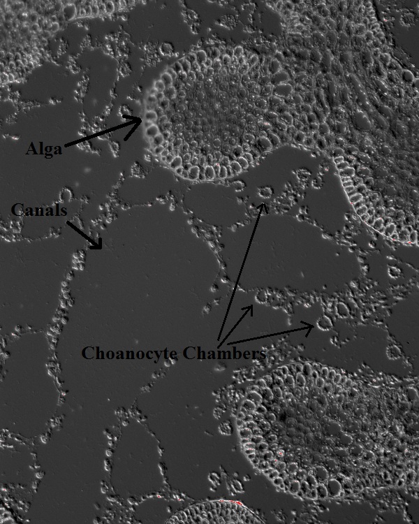 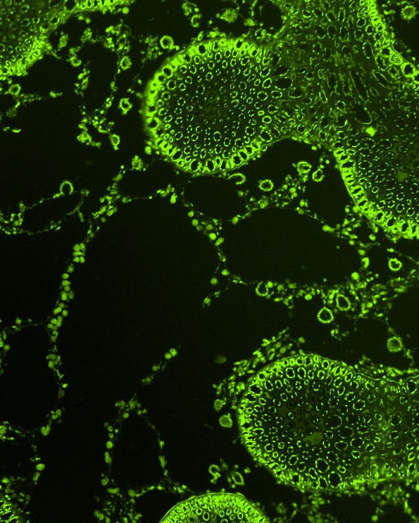
Haliclona/Ceratodictyon (10x): without fluorescence (top); with FITC fluorescence (bottom)
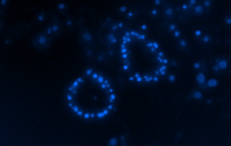 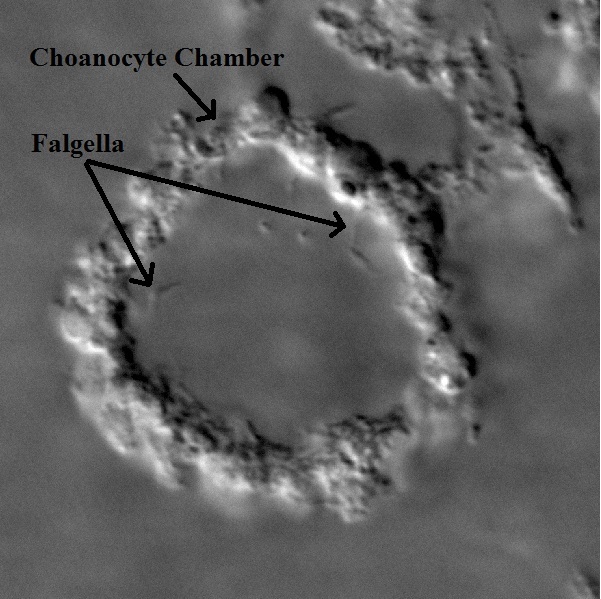
Haliclona/Ceratodictyon
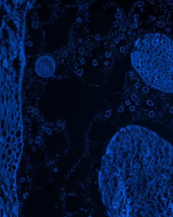
Top (40x): Choanocyte chambers; nuclei stained with DAPI
Bottom (150x): Choanocyte chamber with flagella
Fusion
In Haliclona cymaeformis it has been observed that branches of individual clumps fuse together when they come into contact with one another (Trautman et al., 2003). The actual fusion is initiated after 5-10 days after two individuals got in contact. At this contact point, sponge tissue starts to build a thin “bridge” and after 3-5 days, the fusion of the algal tissue follows. In general, the fusion is completed after 20-25 days after the first contact was made. Once the fusion is completed, it is impossible to tell the branches that formerly belonged to two different individuals apart, not even with a microscope. But these fusions are not always successful. Algal fusion does not necessarily occur after the sponge tissue has built a thin bridge. I some cases, the bridge lasts up to 3 weeks before the sponge tissue is retreated. (Trautman et al., 2003)
|
|